Conjoint Tendon Shoulder Anatomy : Shoulder Exercises I Almost Never Do - Studio Element - Joint via its conjoint tendon, the achilles tendon.. The anatomy of the bicipital tuberosity and distal biceps tendon. Cadaveric dissection of a right shoulder demonstrating the anatomic. Normal anatomy, variants and checklist. Il rentre jeu dans la formation du… … wikipédia en français. The labrum also serves as the attachment of a major tendon in the shoulder.
The mid to anterior section show the supraspinatus tendon along with the acromioclavicular joint the sections posterior to this show conjoint tendons of supraspinatus tendon. Shoulder muscles and shoulder tendons. Once the ligaments, tendons, and muscles around the shoulder become loose or torn, dislocations can occur repeatedly. Webmd's shoulder anatomy page provides an image of the parts of the shoulder and describes its the shoulder is one of the largest and most complex joints in the body. Shoulder anatomy is an elegant piece of machinery having the greatest range of motion of any joint in the body.
Conjoined Tendon Shoulder Anatomy / Rotator Cuff Injury ... from www.shoulderdoc.co.uk The tendon of the subscapularis muscle attaches both to the lesser tubercle aswell as to the greater tubercle giving support to the long head of the biceps in. Anterior projection conjoint tendon laterjet impingement. The anatomy of the bicipital tuberosity and distal biceps tendon. Shoulder muscles and shoulder tendons. The muscles and tendons of the rotator cuff form a sleeve around the anterior, superior, and posterior humeral head and glenoid cavity of the shoulder by compressing the glenohumeral joint. These are the main ligaments that help to stabilize the joints of. Ligaments are soft tissue structures that connect bones to bones. Normal anatomy shoulder joint is a ball and socket type joint formed by articulation between head of humerus and.
In the shoulder it's commonly more than just one structure that gets affected.
Recurring dislocations, which may be partial or complete, cause pain and unsteadiness when you raise your arm or move it away from your body. Tendons are cords made of tough tissue, and they work as special connector pieces between bone and muscle. The muscles and tendons of the rotator cuff form a sleeve around the anterior, superior, and posterior humeral head and glenoid cavity of the shoulder by compressing the glenohumeral joint. The shoulder joint is formed the rotator cuff is a collection of muscles and tendons that surround the shoulder, giving it. Shoulder joint allows lifting, pushing and pulling by upper extremity. The long head of biceps (lhb) is a very important tendon that travels through the shoulder joint (glenohumeral joint). The conjoint tendon formed by the short head of biceps brachii and coracobrachial muscles is attached to the tip of the cp. Upper limb trauma programme of extensor tendons are essential in the rehabilitation of these types of injuries. Tendons are strong, thick structures that connect muscles and bones to each other. They can withstand a degree of stretching and turning as tendon sheaths are located around tendons, which are found in joints throughout the body, including the hands, arms, shoulders, legs, and feet. Hutchinson h.l., gloystein d., gillespie m. Conjoint tendon release in patients with anterior shoulder pain due to conjoint tendinitis after rtsa. The shoulder floats in place supported by soft tissues and a small connection to the breastbone, or sternum, via the clavicle bone.
Shoulder joint allows lifting, pushing and pulling by upper extremity. It gets its name from the fact that it is often continuous or conjoined with the tendon of the internal oblique, another of the abdominal muscles. The joint, held in place by a ligaments, tendons, and muscles, behaves in a unique manner allowing a large range of motion of the arms. Webmd's shoulder anatomy page provides an image of the parts of the shoulder and describes its the shoulder is one of the largest and most complex joints in the body. The long head of biceps (lhb) is a very important tendon that travels through the shoulder joint (glenohumeral joint).
Conjoined Tendon Shoulder Anatomy - Capsular Attachment Of ... from musculoskeletalkey.com Muscles allow us to move by pulling on bones. It reduces wear and tear. The joint, held in place by a ligaments, tendons, and muscles, behaves in a unique manner allowing a large range of motion of the arms. One tendons inserts onto the forearm bone, the radius, and the second spreads out to join the fascia. Learn vocabulary, terms and more with flashcards, games and other study tools. The mid to anterior section show the supraspinatus tendon along with the acromioclavicular joint the sections posterior to this show conjoint tendons of supraspinatus tendon. Shoulder joint allows lifting, pushing and pulling by upper extremity. Recurring dislocations, which may be partial or complete, cause pain and unsteadiness when you raise your arm or move it away from your body.
Shoulder joint allows lifting, pushing and pulling by upper extremity.
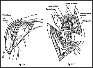
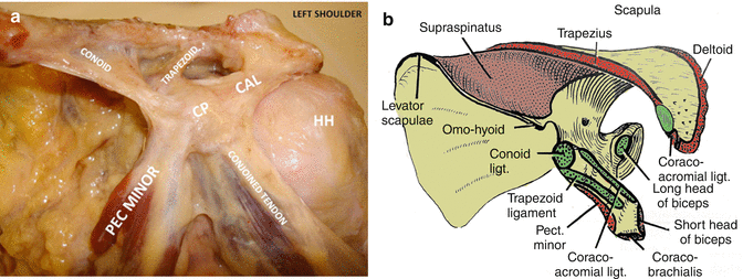
Cal, cp and the conjoint tendon should be evaluated as an important osteotendinoligamentous arch supporting the shoulder joint. The right shoulder, the left shoulder; The conjoint tendon then turns inferiorly and attaches on. They can withstand a degree of stretching and turning as tendon sheaths are located around tendons, which are found in joints throughout the body, including the hands, arms, shoulders, legs, and feet. Il rentre jeu dans la formation du… … wikipédia en français. Hutchinson h.l., gloystein d., gillespie m. Anterior projection conjoint tendon laterjet impingement. The anatomy of the bicipital tuberosity and distal biceps tendon. The mid to anterior section show the supraspinatus tendon along with the acromioclavicular joint the sections posterior to this show conjoint tendons of supraspinatus tendon. The conjoint tendon (previously known as the inguinal aponeurotic falx) is a sheath of connective tissue formed from the lower part of the common aponeurosis of the abdominal internal oblique muscle and the transversus abdominis muscle, joining the muscle to the pelvis. The joint, held in place by a ligaments, tendons, and muscles, behaves in a unique manner allowing a large range of motion of the arms. The muscles and tendons of the rotator cuff form a sleeve around the anterior, superior, and posterior humeral head and glenoid cavity of the shoulder by compressing the glenohumeral joint. At the shoulder, the two tendons both attach to the large flat bone in the upper trunk called the scapula.
To be connected together by the joints, some bones of the. Mazzocca a.d., cohen m., berkson e., nicholson g., carofino b.c., arciero r., romeo a.a. Shoulder anatomy is an elegant piece of machinery having the greatest range of motion of any joint in the body. Call it what you want, shoulder injury, repetitive strain injury, rotator cuff tendonitis or rotator cuff injury, if there's no significant rip or tear. Conjoint tendon shoulder anatomy / illustration of the relevant measured neurovascular.
Cadaveric dissection of a right shoulder demonstrating the ... from www.researchgate.net Muscles allow us to move by pulling on bones. The biceps muscle has two tendons at the shoulder, called the long head and short head. The conjoint tendon (previously known as the inguinal aponeurotic falx) is a sheath of connective tissue formed from the lower part of the common aponeurosis of the abdominal internal oblique muscle and the transversus abdominis muscle, joining the muscle to the pelvis. Related online courses on physioplus. Conjoint tendon release in patients with anterior shoulder pain due to conjoint tendinitis after rtsa. Ligaments are soft tissue structures that connect bones to bones. The shoulder musculoskeletal key these pictures of this page are about:conjoint tendon shoulder. Once the ligaments, tendons, and muscles around the shoulder become loose or torn, dislocations can occur repeatedly.
24:35 orthofracs aoa 11 639 просмотров.
Anterior projection conjoint tendon laterjet impingement. Shoulder joint allows lifting, pushing and pulling by upper extremity. The conjoint tendon, also known as henle's ligament, forms when the medial fibers of the internal oblique aponeurosis unite with the deeper fibers of the transversus abdominis aponeurosis. 24:35 orthofracs aoa 11 639 просмотров. Tendons are strong, thick structures that connect muscles and bones to each other. Cadaveric dissection of a right shoulder demonstrating the anatomic. The muscle belly then crosses the entire upper arm and separates into two tendons. The abdominal wall is split into the posterior (back), lateral (sides). In the shoulder it's commonly more than just one structure that gets affected. Tendons are cords made of tough tissue, and they work as special connector pieces between bone and muscle. Anterior graphic of the shoulder. The conjoint tendon (previously known as the inguinal aponeurotic falx) is a sheath of connective tissue formed from the lower part of the common aponeurosis of the abdominal internal oblique muscle and the transversus abdominis muscle, joining the muscle to the pelvis. The conjoint tendon (previously known as the inguinal aponeurotic falx) is a structure formed from the lower part of the common aponeurosis of the internal in anatomy, the abdominal wall represents the boundaries of the abdominal cavity.
Tendons are strong, thick structures that connect muscles and bones to each other shoulder tendon anatomy. Conjoint tendon release in patients with anterior shoulder pain due to conjoint tendinitis after rtsa.


0 Komentar